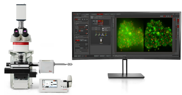
The Leica Cryo CLEM (Correlative Light and Electron Microscopy) imaging system is designed for high-resolution imaging of biological samples in their native, hydrated state. The system is designed to bridge the gap between light and electron microscopy by identifying structures of interest and transferring these coordinates to an EM.
Capabilities include:
- Correlative Light and Electron Microscopy
- Enhanced Visualization: By combining optical and electron microscopy, researchers can gain a comprehensive understanding of sample morphology and internal structures.
- MAPS Software: MAPS software is available to correlate optical images with SEM data, allowing for precise identification of milling locations on samples.
Technical Specifications:
- Filters, Resolution and Objective Setup
- Objective: The system uses the Leica HCX PL APO 50x/0.90 Cryo CLEM objective.
- Numerical Aperture: The objective has a high numerical aperture (NA) of 0.9, allowing for high-resolution imaging.
- Working Distance: It features a low working distance of 0.28 mm.
- Resolution: The maximum resolution is approximately 364 nm to 512 nm depending on the specific configuration.
- Imaging Technique: The system uses widefield fluorescence with THUNDER technology to remove out-of-focus blur (haze), critical for thick cryo-samples.
- Filter Sets: The system is optimized for commonly used fluorophores in cryo-fluorescence: GFP/TRITC, DAPI, and Rhodamine.
Contact: Monica Manginell